Age-related macular degeneration, often called AMD or ARMD, is the leading cause of vision loss and blindness among Americans who are age 65 and older. Because people in this group represent an increasingly larger percentage of the general population, vision loss from macular degeneration is a growing problem.
Age related macular degeneration is a medical condition which usually affects older adults that results in a loss of vision in the center of the visual field (the macula) because of damage to the retina. It occurs in “dry” and “wet” forms. It is a major cause of visual impairment in older adults (>50 years). Macular degeneration can make it difficult or impossible to read or recognize faces, although enough peripheral vision remains to allow other activities of daily life.
The inner layer of the eye is the retina, which contains nerves that communicate sight; behind the retina is the choroid, which contains the blood supply to all three layers of the eye, including the macula (the central part of the retina which surrounds the optic disc). In the dry (nonexudative) form, cellular debris called drusen accumulate between the retina and the choroid, and the retina can become detached. In the wet (exudative) form, which is more severe, blood vessels grow up from the choroid behind the retina, and the retina can also become detached. It can be treated with laser coagulation, and with medication that stops and sometimes reverses the growth of blood vessels.
 Although some macular dystrophies affecting younger individuals are sometimes referred to as macular degeneration, the term generally refers to age-related macular degeneration (AMD or ARMD).
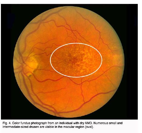
Age-related macular degeneration begins with characteristic yellow deposits in the macula (central area of the retina, which provides detailed central vision) called drusen between the retinal pigment epithelium and the underlying choroid. Most people with these early changes (referred to as age-related maculopathy) have good vision. People with drusen can go on to develop advanced AMD. The risk is considerably higher when the drusen are large and numerous and associated with disturbance in the pigmented cell layer under the macula.
Picture of the fundus showing intermediate age-related macular degeneration
Dry AMD
Central geographic atrophy, the “dry” form of advanced AMD, results from atrophy to the retinal pigment epithelial layer below the retina, which causes vision loss through loss of photoreceptors (rods and cones) in the central part of the eye. No medical or surgical treatment is available for this condition, however vitamin supplements with high doses of antioxidants, lutein and zeaxanthin, have been suggested by the National Eye Institute and others to slow the progression of dry macular degeneration and, in some patients, improve visual acuity.
Beta-carotene was not protective. See also Nutritional supplements (below).
Wet AMD
Neovascular or exudative AMD, the “wet” form of advanced AMD, causes vision loss due to abnormal blood vessel growth (choroidal neovascularization) in the choriocapillaris, through Bruch’s membrane, ultimately leading to blood and protein leakage below the macula. Bleeding, leaking, and scarring from these blood vessels eventually cause irreversible damage to the photoreceptors and rapid vision loss if left untreated.
Until recently, no effective treatments were known for wet macular degeneration. However, new drugs, called anti-angiogenics or anti-VEGF (anti-Vascular Endothelial Growth Factor) agents, can cause regression of the abnormal blood vessels and improvement of vision when injected directly into the vitreous humor of the eye. The injections have to be repeated on a monthly or bi-monthly basis. Examples of these agents include ranibizumab (trade name Lucentis), bevacizumab (trade name Avastin, a close chemical relative of ranibizumab) and pegaptanib (trade name Macugen).
-
 Normal vision (B&W)
 -
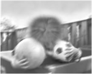 The same view with age-related
macular degeneration (B&W)
The amsler grid, used since 1945, is a grid of horizontal and vertical lines used to monitor a person’s central visual field. The grid was developed by Marc Amsler, a Swiss ophthalmologist. It is a diagnostic tool that aids in the detection of visual disturbances caused by changes in the retina, particularly the macula (e.g. macular degeneration, Epiretinal membrane), as well as the optic nerve and the visual pathway to the brain.
-
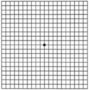 An Amsler grid, as seen
by a person with normal vision. -
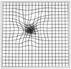 An Amsler grid, as it might be
viewed by a person with age
related macular degeneration.
In the test, the person looks with each eye separately at the small dot in the center of the grid. Patients with macular disease may see wavy lines or some lines may be missing. Amsler grids are supplied by ophthalmologists, optometrists or from web sites, and may be used to test one’s vision at home.
From Wikipedia, the free encyclopedia
Disclaimer
This information is presented for education purposes, to make it easy for you to explore your vision correction options. HOWEVER, no matter how reliable information on this or any website may be, there is no substitute for a professional examination of your eyes and a face-to-face discussion of your unique situation. If you are experiencing any difficulty with your vision, you should schedule an eye examination.